A team of researchers has discovered that the superabsorbent chemical used in disposable diapers can expand brain structures to four and a half times their original size. Referred to as “expansion microscopy”, the discovery would allow scientists to take super-high resolution pictures of healthy and diseased tissue throughout the body without having to improve upon the technological capabilities of today’s high-powered microscopes.

“For centuries, a scientist's ability to look at cells has been constrained by the power of the lenses they used to magnify them. We decided to try something different, and physically magnify the cells themselves,” said Edward Boyden, Ph.D., associate professor of biological engineering and brain and cognitive sciences at the Massachusetts Institute of Technology, Cambridge, and a leader of the study published in Science.
Where the submicroscopic details of cell structures were previously hidden from scientists. Dr. Boyden and his colleagues found that if they linked brain cell proteins to a mesh of sodium polyacrylate and then added water, the mesh swelled and greatly expanded the size of protein complexes without changing or otherwise altering their normal structural arrangement in the cell itself.
“Expansion microscopy is a potential game changer,” said Walter Koroshetz, M.D., acting director of NIH's National Institute of Neurological Disorders and Stroke. “This is the kind of outside- the-box technical innovation that expands the capability of microscopes widely used in the scientific community to explore the fine structure of the nervous system in health and disease.”
To demonstrate the technique’s potential, the team performed experiments on rodent brain slices and cells grown in petri dishes. In both cases, the scientists fixed cell proteins in place with formaldehyde and then stripped the cells of their fatty membranes before treating them with a special labeling molecule that allowed them to visualize protein complexes. Then, before they treated the cells with the acrylate, the team too pictures of parts of the cells using a high powered microscope capable of capturing the finer, more complex details of the cell.
Acrylate was then polymerized to form a mesh and enzymes were used to clear away unlinked proteins; the structures were then expanded with the application of water. Once expansion was complete, pictures of the same locations were taken again, but this time with a lower powered microscope.
Dr. Boyden’s team showed that this expansion microscopy technique improved the second microscope’s ability to resolve the details of the cell’s protein complexes; this includes being able to see the spaces between rows of skeletal microtubule filaments, as well as being able to see the two sides of synapses.
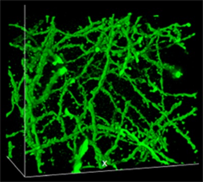
More importantly though, the scientists were able to show that the overall three dimensional protein complexes within a brain cell circuit could be studied in much greater detail, and without having to improve upon the microscope technology itself.
“Our results show that we can scan large chunks of brain tissue with nanoscale precision. We think this can be applied to a variety of tissues and help answer a lot of different questions in science and medicine,” said Dr. Boyden.
Looking ahead, Dr. Boyden and his team plan to test ways of combining their technique with other visualization methods, with the hope of using it to study diseases in human brain tissue.
Via ninds.nih.gov
Advertisement
Learn more about Electronic Products Magazine