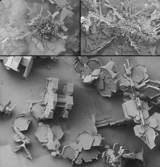
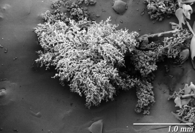
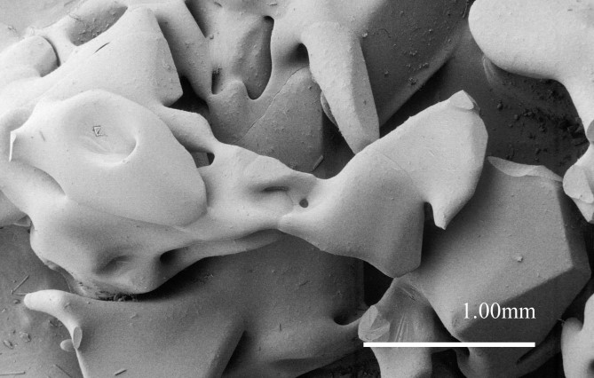
For about one day at a time, those of us living in major cities can overlook our cold and grey misery to bask in the voluptuous coating of freshly laid snow. That momentary respite is short-lived, for the city’s active momentum quickly resumes, transforming all that pretty white powder in slushy grey gunk. But fresh snow doesn’t solely “look pretty” on the macro-level; if you spend a few minutes examining it with an electron microscope, you’ll be treated an array of stunning images adorned in majesty of all things examined by electron microscopes.
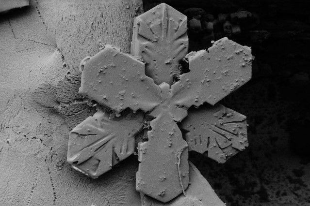
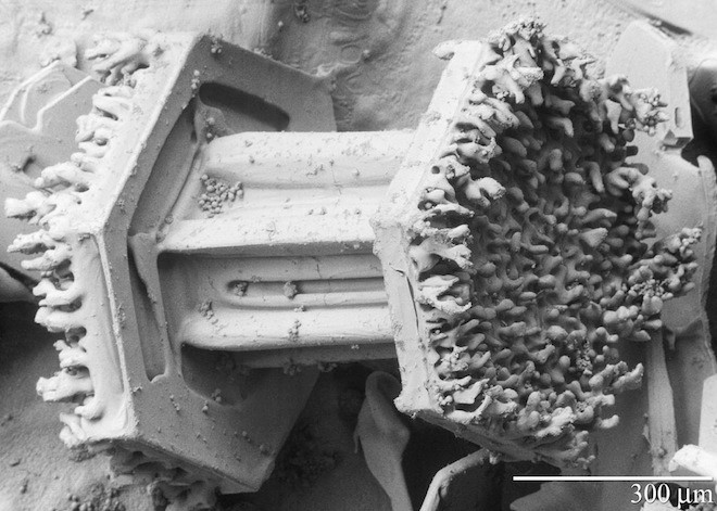
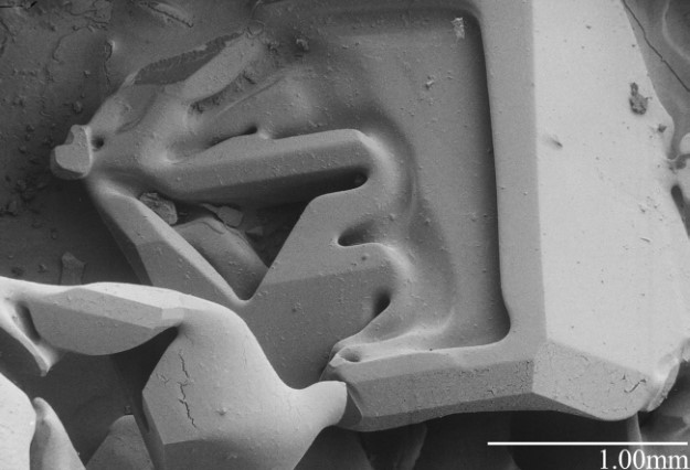
Thanks to the Electron Microscopy Laboratory of the U.S. Department of Agriculture, we can gawk at some fantastic images of snow samples taken from all around the world. Electron microscopes, as the majority of you already know, produce images by blasting objects coated in electrically-conductive material with electrons and observing the rebounded particles. The specific microscope used to produce these images was a Low Temperature Scanning Electron Microscope (LT-SEM).
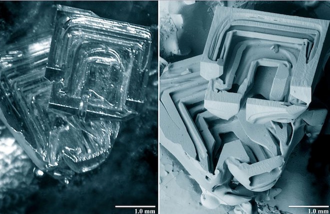
Light microscope versus electron microscope image of hoar crystal from Wyoming snow pit
The studied samples were frozen to a temperature below -321 degrees Fahrenheit before being “sputter coated” with a super-thin layer of platinum in order to make them conductive. Once this was completed, they were shipped to microscopy lab in Beltsville Maryland where the LT-SEM images were snapped and analyzed.
Why does this matter except for our amusement? The research conducted her actually helped create a detailed classification of snow structures that furthered our understanding of water tables, climate change, flooding, and even avalanches.
(All images credit goes to the Electron and Confocal Microscopy Laboratory, Agricultural Research Service, U. S. Department of Agriculture.)
Advertisement
Learn more about Electronic Products Magazine

