Unique considerations in ultrasound beamforming contribute to higher design complexity and better image quality
BY DANIEL KREINDLER
Samplify Systems, Santa Clara, CA
www.samplify.com
Beamforming is a common signal processing technique used to create directional or spatial selectivity of signals sent to or received from an array of sensors or antennae. These arrays can be found in many different devices that transmit and receive either electromagnetic or acoustic waves. Thus, beamforming is employed in such varied applications as radio-astronomy, radar, wireless communications, sonar, seismography, and medical and industrial ultrasound. Ultrasound beamforming is unique among these various applications. In order to achieve high-quality ultrasonic images, received beams must be focused dynamically, and the aperture of the array must be amplitude-weighted (apodized) dynamically as well.
In most beamformer applications, such as in phased-array radar or radio telescope arrays, the distance to the reflector (e.g. an aircraft or a distant galaxy) is large compared to the aperture or length of the antenna or transducer array. In these systems the receiver is primarily concerned with capturing reflections in the farfield. These systems use the array to steer the beam in the needed direction, but can perform static beam focusing with a focal point set at infinity.
Dynamic focusing
In radar or sonar applications, the objects being imaged are typically in the farfield, that is the distance from the sensor array to the object is large compared to the size of the sensor array. In ultrasound beamforming the system must capture both close-in reflections in the nearfield, such as in vascular scans, as well as reflections in the farfield for abdominal or cardiac clinical applications. Consequently, the challenge for ultrasound beamforming systems is to dynamically change the focal point throughout the scan depth from the nearfield to the farfield. This dynamic focusing capability is typical of high quality ultrasound systems and it can provide almost perfect focusing throughout the entire scan depth.
To achieve focusing at different scan depths, multiple foci can be used on the transmit beamformer side. Since any one transmit pulse firing can only focus at a single point, multiple foci require multiple transmit firings per scan line. Since firing the next transmit pulse can only occur after echoes originating from the deepest reflectors have already been received, using multiple transmit foci present the disadvantage of reducing the system’s frame rate. The frame rate is reduced by a factor equal to the number of firings or foci used per scan line.
Like other beamforming applications, the ultrasound receive beamformer can be used to steer the beam by controlling the delay of each element. In addition, by controlling channel to channel delay differences, dynamic focusing can be implemented on the receive beamformer side. These delay differences compensate for propagation delay differences between the focal point and the various elements of the array based on the array geometry. Assuming spherical reflected waves, these delay differences are bigger from targets in the nearfield where the wavefront arriving at the array is more curved, and smaller from those in the farfield where the arriving wavefront is more flat. With dynamic focusing, these focusing delays (added to the steering delays) are not fixed, but rather are a function of time corresponding to the depth or range from which the echoes are being received during the scan-line. Unlike multiple transmit foci, this dynamic focusing on the receive beamformer does not affect frame rates.
Crude implementations of dynamic focusing divide the scan depth into several zones where each has its own focal length. With modern digital beamfomers, the delay resolution can be high enough to allow focusing to track the receive echo depth. This requires ultrasound beamformers to either store in memory or transfer in real time from a processor 1kbyte of data per scan line (assuming 25-cm scan depth with 0.5-mm update increments, and 16-bit data).
Dynamic apodization
Transducer arrays perform sampling in the spatial domain so, as with any sampled data system, steps must be taken to control aliasing effects. Side lobes (or grating lobes when referring to an array) on either side of the main beam can cause image artifacts. Strong reflectors in the beam’s side lobe region can interfere with the receiving of echoes from the targets in the main on-axis beam. Just as time-domain sampling uses an anti-aliasing filter, spatial sampling employs a window function for weighting the transducer elements based on their position in the aperture. This process is called apodization and is accomplished by exciting elements in the array with different voltage amplitudes. The window function used must be selected carefully as there is a tradeoff between the amplitude of the near-in side lobes and the width of the main beam which degrades image resolution. Lateral resolution of an ultrasound system improves as the array aperture decreases in the nearfield and as it increases in the farfield. Thus, as the focal point moves from the nearfield to the farfield, the array aperture is increased by switching on more elements. For optimal apodization, the window function must be continuously scaled to the effective size of the growing aperture. This process is called dynamic apodization.
Most beamforming applications where the targets of interest are in the farfield can use a fixed aperture array along with the fixed focal point. Ultrasound receive beamformers need to dynamically apodize the receive aperture with increasing scan line depth to maintain a constant beam width at the focus for increasing focal lengths. This will maintain a constant lateral resolution throughout the scan line. As with the delay values, apodization weight values at increments in the order of millimeters for each channel are required for dynamic apodization for a scan line. This in turn again requires much memory or high real-time download rates for advanced ultrasound beamformer implementation. Two dimensional (2D) arrays used for 3D imaging require twice as many weight coefficients (for steering in both azimuth and elevation).
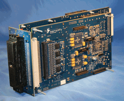
The SMK9130 development kit allows designers to evaluate and use Samplify’s AutoFocus beamformer.
There are many different ultrasound receive beamformer implementations. One unique advanced solution from Samplify Systems integrates the beamformer data path with a calculation engine called AutoFocus. This calculator updates delay and apodization coefficients for each transducer element at 0.5 mm intervals throughout the scan line. This approach supports dynamic focusing and dynamic apodization without the need for any coefficient memory or real-time coefficient downloads from a processor.
Medical ultrasound beamformer designs can be more challenging than those in other fields. In many beamformer applications, like radar, targets are often displayed as single dots on a screen conveying primarily range and velocity information. On the other hand, in modern medical ultrasound systems, beamforming is the first digital signal processing function in a chain that ultimately has to generate highly accurate 2D or 3D images of complex internal human organs, tissue, and blood flow. To deliver the best image quality through improved detail resolution over large depth ranges, it is essential that the beamformer in these systems supports the combination of dynamic focusing and dynamic apodization. ■
Advertisement
Learn more about Samplify Systems