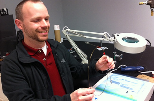
University of Arkansas’ assistant professor of biomedical engineering Timothy Muldoon has developed an endoscopic fiber-optic microscope capable of assisting medical staff in diagnosing the early stages of cancer. Described as “an optical biopsy” by clinicians, Muldoon’s microscope produces high-resolution subcellular images that reveal precancerous cell deformations in real time. Best of all, the microscope is portable, reusable, and, for what it does, inexpensive.
“My dream is to disseminate this technology to a broad scope of medical facilities – hospitals and various clinics, of course, but also to take it into underserved and rural, even remote, areas,” says Muldoon, “its compactness and portability will allow us to do this.” The microscope’s magnifying capabilities enable it to assume dual roles; it may be used as an intraoperative monitoring device to identify the precise location from which to draw a biopsy sample, and it may also be used to observe tumor margins in real time, allowing doctors to verify if they’ve completely removed the tumor.
Professor Muldoon designed and prototyped the microscope while working at the University of Texas’ Cancer Center in Houston. Costing roughly $2,500, the microscope is assembled from thousands of flexible small-caliber fibers that make up a single fiber optic bundle measuring approximately one millimeter in diameter ─ small enough to insert the microscope into the biopsy channel of an endoscope. The probe must be sterilized prior to use and fitted with a topical contrast agent to facilitate the fluorescent imaging.
Once the prototype was completed, Muldoon conducted the first series of field tests while residing at the University of Texas. It was determined that the microscope’s view of the nuclear-to-cyptoplasmic ratio of esophageal cells, a cell behavior leading to precancerous conditions, matched results obtained by the standard histopathological’s examination of biopsied tissue. Currently, Muldoon is collaborating with a surgeon and clinical pathologist at the University of Arkansas to test how the microscope stands up against colon cancer cells.
If the microscope passes medical scrutiny, it could prove extremely useful in monitoring all manner of medical treatments in real-time by directly observing how the cells react. And it will need an official name.
Advertisement
Learn more about Electronic Products Magazine


