A multiyear project has led a team of researchers to a possible major breakthrough in biotechnology — that is, they’ve created a small, intracranial robot that can be used to effectively suck up and eat away at brain tumors.

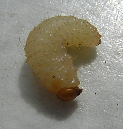
J. Marc Simard, a neurosurgery professor at the University of Maryland School of Medicine, led the team’s research. He described the maggot-like device as being shaped somewhat like a mechanical finger, complete with multiple joints that give the mini-bot a range of motions.

Initial prototype for the minimally invasive neurosurgical intracranial robot.
At the tip of the device is an electrocautery tool, which uses electricity to heat up and destroys tumors. It also includes a suction tube to suck out left-behind debris.
“The idea was to have a device that’s small but that can do all the work a surgeon normally does,” said Simard. “You could place this small robotic device inside a tumor and have it work its way around from within, removing pieces of diseased tissue.”
Speaking of placement, the robot gets inserted into the patient’s brain, at which point the individual is placed inside an MRI scanner. Doing this, the group discovered, provided the doctor with unprecedented views of otherwise hard-to-see tumors. As for controlling the bot, the doctor sits in a nearby room and steers the device via a remote-controlled system.
Development of the remote control for this technology was no easy feat, according to Jaydev Desai, the team’s mechanical engineer. He explained that the most challenging part was designing a robot that can be controlled inside the magnetic field of an MRI.
You see, robots are typically controlled via electromagnetic motors. You can’t do this inside an MRI machine — besides being magnetic, the motors create image distortion, clouding the surgeon’s on-screen field of vision and making it impossible to perform the task.
Hydraulic systems were considered as an alternative but quickly dismissed due to the risk of fluid leakage.
So, to overcome this engineering hurdle, Desai used shape memory alloy (SMA) to control the robot’s movement. SMA is a material that alters its shape in response to changes in temperature.
In the most recent prototype, a system of cables, pulleys, and SMA springs were used and proved to be an improvement over the previous model, which caused some image distortion.

Newest prototype for the minimally invasive neurosurgical intracranial robot, which uses a system of pulleys and springs to move the robot about.
Simard explained that he was inspired to embark upon this project after watching a TV show in which plastic surgeons used sterile maggots to remove damaged / dead tissue from a patient.
“Here you had a natural system that recognized bad from good and good from bad,” he said. “In other words, the maggots removed all the bad stuff and left all the good stuff alone and they’re really small. I thought, if you had something equivalent to that to remove a brain tumor that would be an absolute home run.”
What’s more, operating within an MRI machine not only allows the surgeon to see the unseeable, it also allows for the continued monitoring of the tumor’s borders throughout the procedure.
The entire approach provides several major advantages, according to Steve Krosnick, M.D., a program director at the National Institute of Biomedical Imaging and Bioengineering.
“Unlike pre-operative MRI or intermittent MRI, which requires interruption of the surgical procedure, real-time intra-operative MRI offers rapid delineation of normal tissue from tumor while accounting for brain shifts that occur during surgery.”
Simard adds, “When we’re operating in a conventional way, we get an MRI on a patient before we do the surgery, and we use landmarks that can either be affixed to the scalp or are part of the skull to know where we are within the patient’s brain.” He went on: “But when the surgeon gets in there and starts to remove the tumor, the tissues shift around so that now the boundaries that were well-established when everything was in place don’t exist anymore, and you’re confronted once again with having to distinguish normal brain from tumor. This is very difficult for a surgeon using direct vision, but with MRI, the ability to discriminate tumor from non-tumor is much more powerful.”
For now, the maggot-bot is still in development, and the team will spend a good deal of time trying to make the picture clearer and the bot smaller. When the device is deemed ready for action, it’ll have to go through an extensive series of clinical tests before it shows up at any local hospital.
Story via: nibib.nih.gov
Advertisement


