Image via MIT
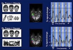
MIT neuroscientists at the McGovern Institute for Brain Research have developed a technique that looks at how and where humans process information in their brain. To test this concept, the scientists scanned peoples’ brains while they viewed a series of images. While scanning the brain, the scientists saw where the mental connection was occurring, and could see the brain while it recognized an object, categorized it, and then processed it as an entity.
Scientists at MIT’s Computer Science and Artificial Intelligence Laboratory (CSAIL) have been determining how the brain interprets the “when” and “where” of images.
Before researchers began working on this project, location or timing in the brain’s activity were examined at high resolution. Both of these processes could not be simultaneously observed because of the prior lack of technological advancements. The functional magnetic resonance imaging (fMRI) scan analyzes the brain’s blood flow while participating in activities. This form of scanning is not nearly fast enough to keep up with the brain’s millisecond-to-millisecond functionality.
Magnetoencephalography (MEG) is another form of imaging used by the team at MIT. MEG uses head-encircling sensors to observe the magnetic fields emitted by the brain’s neuronal activity. Through these measurements, the sensors paint a highly precise picture of brain activity over a given period in time.

Image of an fMRI brain scan via MIT
Researchers then combined the time and location data gathered from the scanners. Representational similarity analysis, a computational technique, is used to determine how the brain processes two similar objects (like two faces) that will generate related signals in fMRI and MEG. Before using this technology on humans, monkeys’ neuronal electrical activity was measured.
In order to receive accurate test results, 16 humans were scanned voluntarily after looking at 92 images, spending half a second on each image. The test subjects received the exam many times, going twice in an fMRI scanner and twice in an MEG scanner.
The scientists measured the data and determined a timeline that shows the brain’s recognition pathway. All visuals shown to the test subjects entered the brain’s primary visual cortex (V1), recognizing things like shapes. Next, the brain’s inferotemporal cortex received the information where the object was identified within 120 milliseconds. All images were categorized into groups within the brain (for example, if the object was a plant or animal).
Currently, the team at MIT is using representational similarity analysis to observe computer models of vision and exactly how accurate they are in reference to how human vision works.
Moving forward, the experts at MIT will study how the brain processes motor, sensory, and verbal information. This research will assist in potentially determining the cause for memory disorders, paralysis, or dyslexia.
Story via Phys.org
Advertisement
Learn more about Electronic Products Magazine


