Physicists from the University of Twente in Enschede, the Netherlands, are refining a new form of imagery that uses visible light waves to perform the same function as x-rays, only without the lingering radiation. So far, the technique is able to penetrate solid objects like paint and mouse flesh, creating the hope that it will someday replace x-rays, or potentially allow doctors to remove tumors using lasers, rather than surgery.
The initial experiments leveraged a technique from astronomy called “adaptive optics,” which uses computer algorithms to calculate the amount of image’s distortion based on atmospheric conditions. This technique was then adjusted to opaque objects by shining a laser through a spatial light modulator, a device that enacts spatially varying modulation on a beam of light, allowing them to delay segments of the beam before striking the opaque object. Next, a sensor on the other side of the object determines the source of the scattered light and stitch together a coherent, unified image.
To the surprise of the physicists involved, Allard Mosk and Ivo Vellekoop, the first few experiments generated a concentrated pinprick of light one hundred times brighter than scattered light. “This just doesn't happen on the first day of your experiment,” exclaims Mosk. “We thought we'd made a mistake and there must be a hole in our slide letting the light through!”
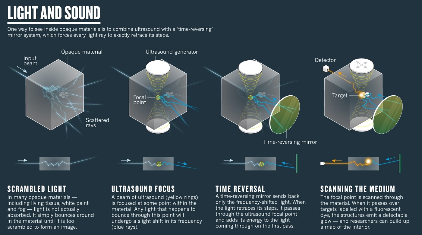
Inspired by these results, a different team of physicists adjusted the technique to incorporate ultrasonic waves, shifting the laser light frequency. The frequency-shifted rays were then bounced back through the object using a “time-reversing mirror,” adding to the energy of the beam from the first pass to create a “torch inside the wall” — a section of concentrated ultrasound focus and high radiative intensity. This permitted the team to image a fluorescent bead, measuring only a single micrometer across, centered between two opaque layers.
The latest breakthrough came from a yet another team of researchers, this one led by Sylvain Gigan, a physicist at the Kastler Brossel Laboratory in Paris, have managed to apply the technique in imaging the ear of a living mouse back in 2014, the first attempt at living flesh. But of course, moving from a imaging mouse ear to human flesh will take years of work, and require a mode of dealing with tissue that moves or stretches. Nonetheless, visible-light images taken from deep within the body may eventually eliminate the need for intrusive biopsies.
Source Nature
Advertisement
Learn more about Electronic Products Magazine