Researchers develop X-ray machine that fits in your palm
BY NICOLETTE EMMINO
Soon you may be able to fit an X-ray machine in the palm of your hand. An inexpensive radiation source the size of a stick of gum has been developed by University of Missouri researchers to make handheld scanners or tricorders as they are being called, possible. A prototype for these scanners could be ready in about three years.

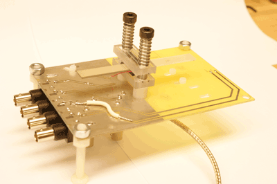
The compact radiation source developed by researchers at the University of Missouri. (Image via Peter Norgard, University of Missouri)
So why would we want portable handheld X-ray machines?
“Currently, X-ray machines are huge and require tremendous amounts of electricity,” said Scott Kovaleski, associate professor of electrical and computer engineering at the University of Missouri. “The cell-phone-sized device could improve medical services in remote and impoverished regions and reduce health care expenses everywhere.”
Tiny X-ray generators would be safer to use. In dentist offices, they could be used to capture images inside of a patient’s mouth in order to shoot the rays outward and reduce exposure to the head.
Portable scanners could also be used to search cargoes at border crossings to improve security and reduce costs. Since they require little energy, they could potentially be used on space rovers as well.
How it works
The device uses a crystal that produces more than 100,000 V of electricity from only 10 V of electrical input with low power consumption. Since the crystal requires so little power, it could eventually be fueled by batteries. The crystal is composed of a material called lithium niobate and amplifies the input voltage by means of piezoelectricity.“The device is about 10 cm long x 1 cm wide x 1.5 mm thick. When we apply a 40000 Hertz, 10 Volt, AC voltage through the thickness, an inverse piezoelectric effect is induced, causing the crystal to expand and shrink through the thickness,” said Kovaleski.At the high voltage end are attached bits of sharp metal called field emitters which emit electrons in the strong electric field produced at their sharp tips. Once these electrons are accelerated toward a target, the are converted into X-rays.“Using an optical camera analogy, our x-ray source acts like a flash illuminated the subject that might be imaged onto film or a digital image capture device,” said Kovaleski.
Koleski does not envision that his newly developed accelerator would be used only in X-ray machines. “Our device is perfectly harmless until energized, and even then it causes relatively low exposures to radiation,” said Kovaleski.
He sees that it could also replace radioactive materials such as radioisotopes used in drilling for oil or in other industrial and scientific operations. These radioisotopes could be replaced by a safer source of radiation that could be turned off in case of emergency. ■
Check out the research paper below for more information.
Advertisement
Learn more about Electronic Products Magazine


