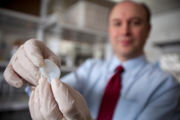
Researchers from China and the US have used 3D-printing techniques to create a 3D cancer “tumor,” a model that could provide more realistic insight into the disease during cancer and drug studies. 
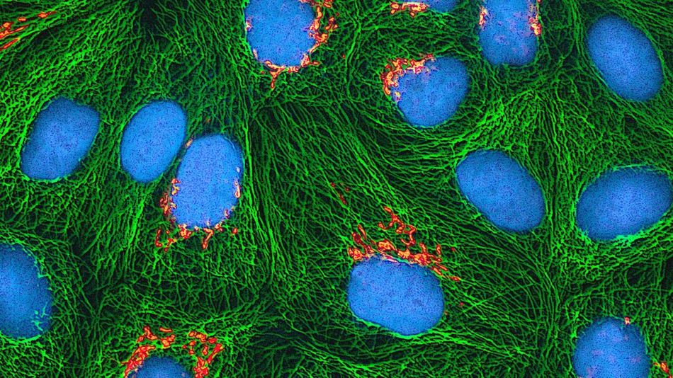
Led by Philadelphia Drexel University and China Tsinghua University professor Wei Sun, the researchers used mixed gelatin, fibrous proteins and HeLa cervical cancer cells to create their structure. HeLa cells, as they were originally taken from a 1951 cancer patient named Henrietta Lacks, are often referred to by scientists as the “immortal cell line,” and can multiply indefinitely. The team fed these components into a 3D-printer of their own design, which produced a tumor-like grid structure of cancer cells.
Thanks to their use of fibrous proteins, the 10 millimeter wide and 2 millimeters tall grid resembled the extracellular matrix of a cancer tumor, before the team allowed the cells to grow on their own. After five days of independent growth, the grid took on a spherical shape, which itself grew for three more days. The team had to create their structure within certain parameters to ensure the majority of the cells survived the printing process itself, as the actual mechanics of printing could cause damage to the cells.

Unlike the typical 2D models of cancer tumors one would typically see in a lab or during cancer studies, the 3D tumor produced by the team provides a much closer look at how cancer works. The tumor’s additional dimension reveals information about cancer cells’ shape, proliferation, and even gene and protein expression that a 2D model would not be able to show.
The team’s results were published in the journal Biofabrication
Source Mashable
Advertisement


